orimtech
tomography for large animals

RADIOGRAPHY, TOMOGRAPHY, TOMOSYNTHESIS
AND FLUOROSCOPY IMAGING CAPABILITIES INTEGRATED IN A SINGLE SYSTEM

IPS Medical is steadfastly advancing the development of new technologies, with the goal of providing ever more advanced and high-performing products to the international market.
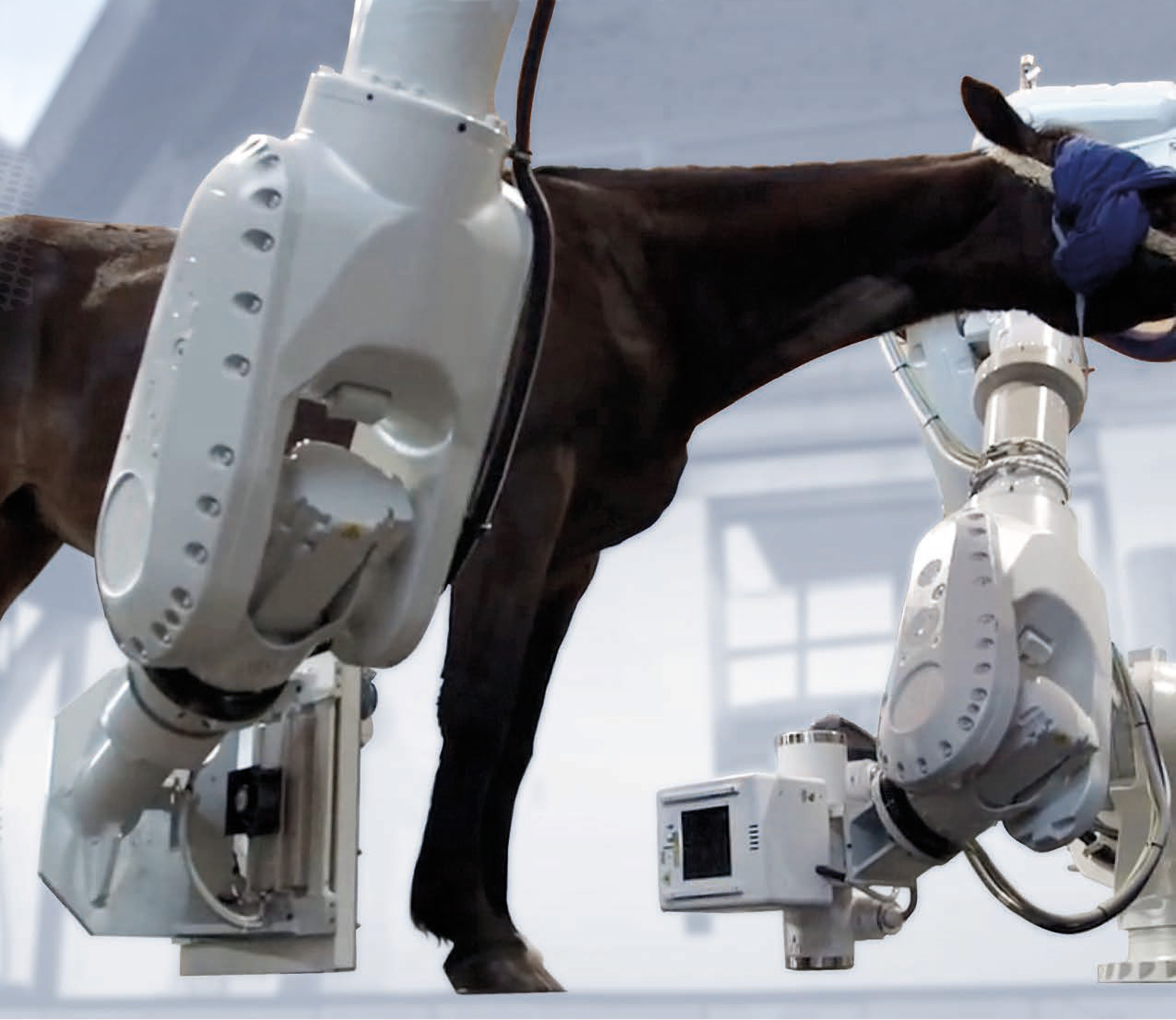
In this spirit, we have established a prestigious partnership with the American company Orimtech for the distribution and support of the EDAMIS system: a revolutionary and unique solution that integrates tomography, tomosynthesis, digital radiography, and fluoroscopy into a single device, specifically designed for large animals such as horses and camels.
EDAMIS represents the future of veterinary diagnostics, combining versatility, precision, and unprecedented technological innovation.
- Common features
- CBCT specifications
- Fluoro specifications
- Radiography specifications
- Tomosynthesis specifications
- Modalities: Radiography, Fluoroscopy, Cone Beam CT, Tomosynthesis.
- Available X-Ray technique factors are defined by the integrated X-Ray generator, tube, tube assembly, and detector. Currently the supported system configuration are based on X-Ray Swiss Genesis (Josef Betschart AG, Switzerland) or CPI Indico IQ 100 (Canada ) generator and Varex XRD 4343 RF (Germany) or iRay Mercu1717V (China) panels. COMET generators are also supported.
- Collimation: 4 blade PC-controlled automatic collimator
- Acquisition: Orimtech proprietary acquisition/synchronization system
- Object/patient database
- DICOM support; PACS compatible
- Common advantage: high flexibility in selection of patient-system orientation, source-detector distances and system scanning trajectories for all modalities – fully defined by specific robot models.
- X-Ray factors, techniques, and frame rate: defined by the integrated X-Ray generator, tube, detector.
- Scanned object positioning/orientation: arbitrary (within the system mechanical accessibility), up to 12 User-predefined scanning trajectory protocols
- FOV adjustment and scouting: collimator light and laser, joystick controlled system motion, linear X-Ray scanning in a single or dual orthogonal planes scout scans to allow for further User-PC interactions to definine area of interest for a scan.
- Scan angle: 210 degrees
- Field of view: D240mm x 240mm one slab; with optional 2 stitched slabs with up to D240mm x 420mm of total coverage, depending on tube/detector distance configuration.
- Spatial resolution: 250 micron (mostly defined by a tube focal spot)
- Acquisition technique: pulsed, up to 25 FPS.
- Single slab scanning time: 10-60 sec depending on the X-Ray dosage
- Supported scanning trajectories: circular and non-circular; built-in trajectory registration for the best spatial resolution.
- Ability to scan solid moving objects (motion artifact mitigation): supported by integrated Qualysis visual motion tracking subsystem.
- X-Ray technique modulation during the scanning process for the optimal patient dose utilization: supported
- CT reconstruction engine: AVRG with support of scatter, beam-hardening and metal artifact reduction.
- HU accuracy: +/-25 HU on CTP486 section (CATPHAN)
3D image viewer: Orimtech MPR proprietary, Fovia HDVR is an option
- X-Ray factors, techniques, and frame rate: defined by the integrated X-Ray generator, tube, detector.
- Automatic brightness control: hardware and software supported
- Real-time display and playback: supported with Orimtech proprietary dynamic contrast enhancement function
- FOV position control: joystick , PC keyboard and touch screen
- Subtractive fluoroscopy techniques (for contrast studies): supported
- X-Ray factors, techniques: defined by the integrated X-Ray generator, tube, detector.
- Spatial resolution: mostly defined by X-Ray panel; approximately 5LP for XRD 4343 RF
- Imaged object positioning/orientation: arbitrary, within the system mechanical accessibility
- Source-detector distance: arbitrary, within the system mechanical accessibility
- FOV adjustment: collimator light and laser, joystick controlled system motion.
- DR image processing library: Orimtech proprietary, CVIE (Context Vision)-ready
- X-Ray factors, techniques, and frame rate: defined by the integrated X-Ray generator, tube, detector.
- Scanned object positioning/orientation: arbitrary (within the system mechanical accessibility) , up to 8 User-predefined scanning trajectory protocols.
- FOV adjustment and scouting: collimator light and laser, joystick controlled system motion, linear X-Ray scanning in a single or dual orthogonal planes with further User-PC interactions for defining the scanned area.
- Scan angle: 15-90 degrees for angular tomosynthesis examinations, 360 degrees for circular motion.
- Field if view: D240mm x 240mm
- Spatial resolution: 200 micron (mostly defined by a tube focal spot)
- Acquisition technique: pulsed, up to 25 FPS.
- Scanning time: 4-20 sec
- Supported scanning trajectories: circular and non-circular; built-in trajectory registration for the best spatial resolution.
- Ability to scan solid moving objects (motion artifact mitigation): supported by integrated Qualysis visual motion tracking subsystem.
- X-Ray technique modulation during the scanning process for the optimal patient dose utilization: supported
- Tomosynthesis reconstruction engine: AVRG (Orimtech)
- 3D image viewer: Orimtech proprietary

The use of large-scale industrial robots, powerful X-ray source (100 kilowatts), and X-ray detectors from the world’s best manufacturers.

Specially designed proprietary algorithms for processing digital radiography and fluoroscopic images. Compensation of the high-level of radiation absorption. The ability of performing tomosynthesis and fluoro studies of spine, including the immediate vicinity of the shoulder and pelvis areas.

Registration and compensation of animal motion to obtain high spatial resolution images. No need for deep anesthesia.

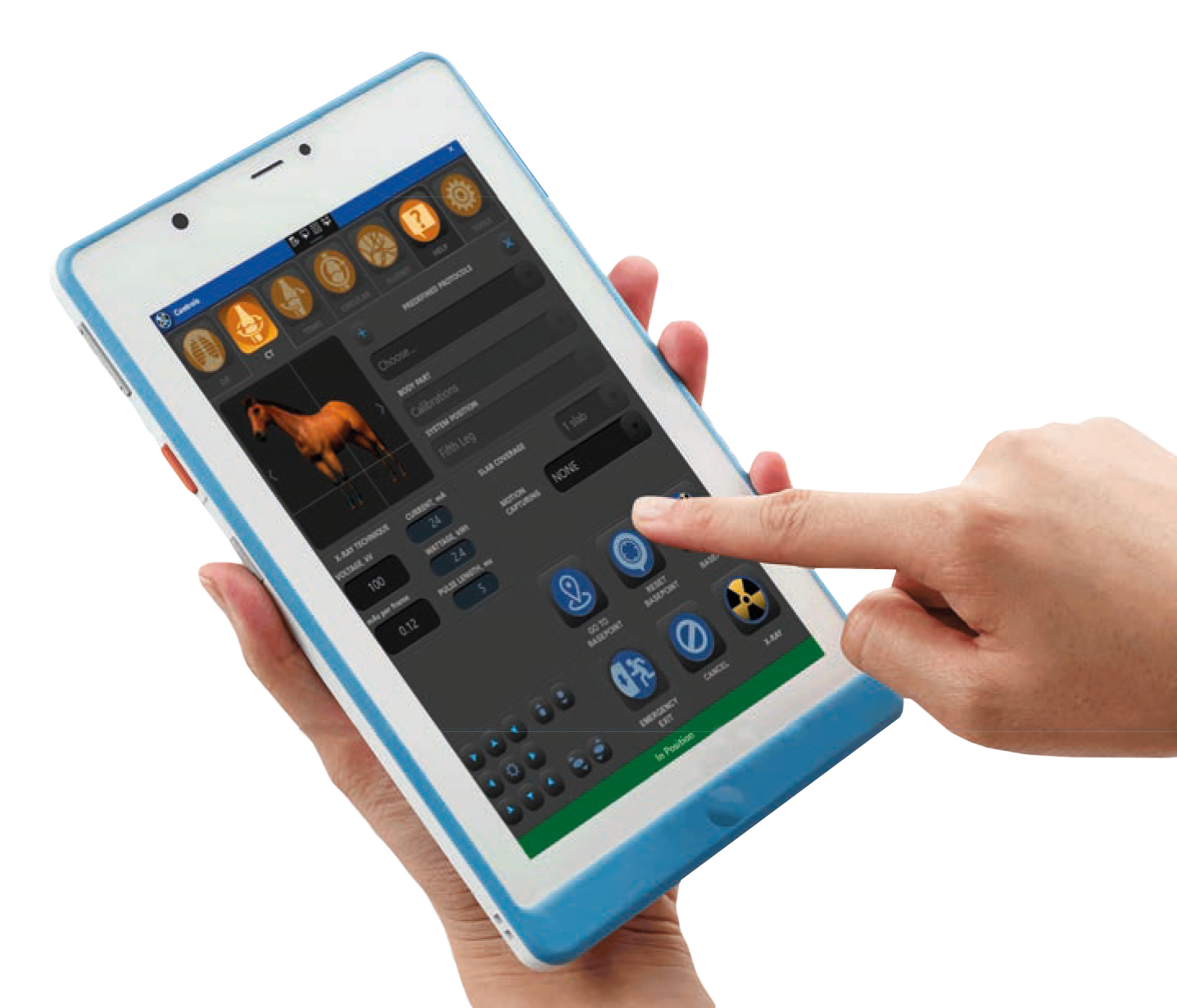
Easy to manage: controlling robots and radiation techniques by a consumer mobile device in the immediate vicinity of the patient. Built-in video recording of the study.
do you need help?
rely on our service department
Contact our specialists for remote technical support of veterinary radiology and diagnostics.
contact us
For information about our products and services
I.P.S. Medical Srl
Via dell’Agricoltura 22/24, 37012 – Bussolengo (VR), ITALY
Phone +39 045 67 02 927 | Mail sales@ipsxray.com
Business hours
Mon – Fri 08.30 – 12.30 / 13.30 – 17.30 (UTC+1)
Technical assistance
Phone +39 327 5315925 | Mail engineering@ipsxray.com
Follow us